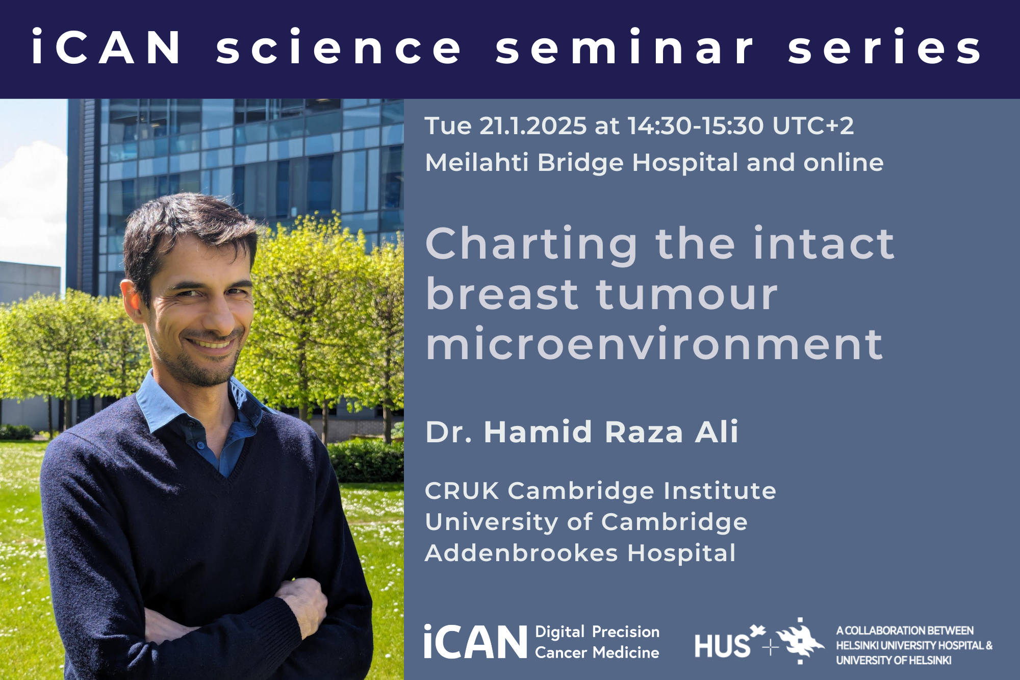
iCAN science seminar on Charting the intact breast tumour microenvironment with Dr. Ali
The diagnosis and treatment of breast cancer continues to rely on decades-old techniques in traditional histopathology. Immunotherapy has proved effective among some patients but not others, and this variation is poorly explained by traditional assays. Using imaging mass cytometry – a technique that couples antibodies conjugated to rare earth metal reporters and time-of-flight mass spectrometry to infer epitope abundance at subcellular resolution – Dr. Ali’s group has shown that the complexity of the TME can be reliably enumerated in situ and used to predict response in a large, randomised trial of neoadjuvant immunotherapy in triple-negative breast cancer. Moreover, the group’s research show how immunotherapy remodels the TME, and how resistant cancer cells endure treatment by analysing serial samples collected over the treatment course. In his talk Dr. Ali will share their results and offer some insights on the wider implications for spatial cancer biology.
To hear more, join us at iCAN Science seminar
Charting the intact breast tumour microenvironment with Dr. Hamid Raza Ali
on Tue 21.1.2025 at 14:30-15:30 (UTC+2)
at Meilahti Bridge Hospital, lecture room Ullanlinna
or on Zoom (Meeting ID: 648 0498 6682 Passcode: 366409)
If you wish to meet with Dr. Ali during his visit, please contact the host, Prof. Anniina Färkkilä (anniina.farkkila@helsinki.fi)
For students: if you are interested in joining a student lunch prior to the seminar, please contact Iga Niemiec (iga.niemiec@helsinki.fi) for more information.
Minibio of the speaker
Dr. Ali is Leader of the Systems Cancer Pathology group, based at CRUK Cambridge Institute, University of Cambridge, Associate Director for Clinical Academic Training , and an Honorary Consultant Pathologist at Addenbrookes hospital. He read medicine in Cardiff and began his training as a pathologist there before moving to Cambridge to undertake a PhD in the quantitative pathology and genomics of breast cancer, under the supervision of Prof Carlos Caldas. He completed his specialist clinical training in Cambridge as an NIHR Clinical Lecturer before moving to University of Zurich. In Zurich, he worked under the supervision of Prof Bernd Bodenmiller, inventor of imaging mass cytometry. There, he conducted research using highly multiplexed epitope-based tissue imaging to understand the principles of spatial organisation that characterise breast tumour ecosystems. His group uses imaging mass cytometry to understand the spatial dynamics of breast cancer through disease progression and treatment, in order to identify adaptations and biomarkers associated with relapse and response.
The seminar is open to all interested in the topic. Come early for a cup of coffee, pulla, and great discussions in excellent company!
Welcome!


